Cause of Clefts
What is a cleft lip and palate? A cleft lip and palate occurs when there is a partial or complete gap in the formation of the upper lip, the gum of the upper jaw and the roof of the mouth. That gap occurs because of a failure of the various parts of the upper lip, the upper jaw and the roof of the mouth to join properly during the formation of the baby in the early weeks of pregnancy in the womb. The exact nature of the cleft in any individual baby depends upon which of those parts fail to join together.
Normally the upper lip, the upper jaw and the roof of the mouth is formed in three separate parts. Those parts join together when the foetus in the womb is between 7 and 12 weeks old. But under certain circumstances, of which we are unaware, those parts may fail to join together. If that should occur, the result is a partial or a complete cleft. The cleft may involve the lip alone, the lip and the gum together or in its complete form; the lip, the gum and the roof of the mouth. Alternatively the cleft may only involve the roof of the mouth with no visible gap in the lip or the gum.
In Australia, a cleft occurs randomly in approximately 1 in 800 live births. The risk, however, is increased where there is a previous family history. For example, if one parent has a cleft, the risk of that couple having a baby with a cleft is 1 in 50 (2%). If that same couple does in fact have one baby with a cleft and wishes to have another child, the risk increases to 1 in 6 (15%). If a couple in which neither parent has a cleft have a baby with a cleft and wish to have another child, then the risk of a second child with a cleft is 1 in 20 (5%).
The majority of children with cleft lip and palate have no other congenital abnormalities. However, there are a variety of rare syndromes where a cleft lip and palate can be part of a number of other congenital abnormalities. There are many such syndromes but each of them is really quite rare.
As far as we are aware there are no chemicals or drugs which can result in a cleft lip and palate
General Outline of Treatment
-
Pre-natal Diagnosis
Increasingly with advances in ultrasound technology, the diagnosis of cleft lip and palate may be made before the birth of the baby. The earliest stage at which cleft lip and palate can be diagnosed is at about the 18th to 20th week of pregnancy and members of the Cleft Lip and Palate Team are sometimes invited to counsel parents where a pre-natal diagnosis of clefting has been made. This can be very helpful to those parents in preparing them for the birth of their baby and in discussing at an early stage the treatment.
-
Treatment Management
Treatment and management of any child’s cleft depends on the extent and severity of the cleft. In the most severe forms of complete cleft lip and palate, all members of the cleft lip and palate team will be involved at some stage during the child’s growth and development. On the other hand, where there is a cleft of the lip alone, or alternatively, a cleft of the palate alone, then the treatment program will not involve all of the members but only those appropriate to the child’s type of cleft.
-
Neonatal Feeding
Those babies born with a cleft of the palate will inevitably have some difficulty in early feeding. The normal sucking mechanism requires an intact roof of the mouth (palate) to ensure that the baby can suck properly. Where there is a gap in the roof of the mouth it can be extremely difficult for the baby to successfully breast feed and alternative methods of feeding will need to be introduced. This usually involves the use of a compressible feeding bottle with a modified nipple. Midwives who have special expertise in this area can help mothers to manage with feeding. In extreme cases a baby may require a naso-gastric (stomach) tube to assist with feeding in the early stages.
-
Surgical Treatment
The particular operation needed for the baby varies and depends upon the specific nature of the cleft. Details of the surgery and the timing of operations are outlined further in this booklet.
-
Hearing
All children with a cleft of the palate have the potential for impaired hearing. This is due to middle ear dysfunction, which will be exp∏lained further in the booklet, and these children are carefully monitored to evaluate their hearing.
-
Speech
As with hearing, all children with a cleft of the palate need to be monitored very closely to ensure that their speech develops normally. Approximately 80% of children with a cleft palate will proceed to normal speech with approximately 20% requiring speech therapy. Arrangements will be made through the Cleft Lip and Palate Clinic to ensure all children receive regular speech appointments.
-
Dental and Orthodontic Care
All children seen and managed through the Princess Margaret Hospital Cleft Lip and Palate Clinic are entitled to have their dental care undertaken at Princess Margaret Hospital. Those with a cleft of the lip alone and a cleft of the palate alone may not require much dental treatment, but all children who have a cleft of the gum will require specialist dental care. This will include normal restorative dentistry, orthodontic treatment and in some cases, specialised dental treatment both before and after the child has all its adult teeth.
Cleft of the Lip
-
Embryology
The upper lip is formed in three parts. There is a relatively small central part to the upper lip and two relatively larger parts on either side. It is possible therefore to have a cleft of the lip, which is either on the right or the left (unilateral), or alternatively, it is possible to have a cleft which affects the right and the left (bilateral). Essentially the cause is the same for all of these variations, namely a failure of one or more of the three elements of the upper lip to join together when the foetus in the womb is 7 to 9 weeks old. Occasionally with the cleft lip alone, the separation can include a small notch in the gum only and if that does occur it may ultimately result in the teeth in the region of that notch erupting slightly out of alignment.
-
Feeding
There is generally no problem at all in a cleft lip baby with respect to feeding. Providing the palate is intact, there should be no difficulty in this child feeding normally at the breast, or alternatively, with a normal bottle.
-
Neonatal Dental Treatment
In children with a cleft lip alone, there is no need for neonatal dental treatment and no need for any dental plates.
-
Surgery
The cleft of the lip is normally repaired at about three months of age. Under certain circumstances it is possible to undertake the surgery earlier, but generally that is discouraged to allow the baby to grow and be a little bit more robust for the operation. At the time of the operation, the baby will be admitted to Hospital for approximately five days and the operation itself usually takes about 1 and a half hours. During surgery, the edges of the cleft lip are incised in a zigzag manner and then carefully stitched together, paying particular attention to the muscles and skin of the lip. At that same time, surgery is done to the nose, as often in children with a cleft lip there is some distortion and asymmetry of the nose. This is best corrected at the time the lip is being repaired. It may be necessary to cease breastfeeding for approximately one week following the operation and during that time the baby will need to be fed milk with a spoon. Once all of the stitches have been removed at about five days post surgery, the child will be ready to return home. This operation usually makes a dramatic change to the appearance of the baby and of course it does result in a small scar on the upper lip which will slowly and steadily fade with time. Nonetheless, the child will always have a permanent scar on the upper lip. In some instances, if the scar remains noticeable, a minor corrective procedure can be undertaken just prior to going to school.
-
Hearing
As a general rule, children with a cleft of the lip only will have no increased risk of middle ear dysfunction, and therefore no greater risk of hearing problems in comparison to normal children.
-
Speech
Unless the child with a cleft of the lip also has a cleft palate, then there would be an expectation of normal speech.
-
Follow Up
All children treated in the Cleft Lip and Palate Clinic are followed up at regular intervals to ensure growth and development is normal. Moreover, these visits give us the opportunity to measure and record progress, ensuring correct and appropriate treatment is undertaken.
Cleft Lip of the Palate
-
The Embryo
When the foetus is 12 weeks old the upper lip, the gum and the roof of the mouth are formed. The formation of these structures is in three parts with each of the parts joining together to form a single symmetrical block of tissue which includes the upper lip, the gum and the associated teeth and the roof of the mouth. In those children who are born with a cleft of the lip and palate, there is a failure in part or in total of the three elements to join together – we call this a cleft. The cleft may occur on the left hand side, the right hand side or in its most severe form involves both sides. In the former two situations, we have what is called a unilateral cleft of the lip and palate, whilst when the failure to join occurs on both sides we have what is called a bilateral cleft of the lip and palate. Whether the cleft is unilateral or bilateral, there is an opening between the mouth cavity and the nasal cavity in those children with a complete cleft of the lip and palate. Why this phenomenon occurs we do not know but there is much that can be done to correct this congenital deformity.
-
Feeding
As one might expect, where there is no intact roof of the mouth it is extremely difficult, if not impossible, for a baby to suck normally. This generally makes breastfeeding impossible, but where the cleft is incomplete or where it is not particularly wide, occasionally it may be possible for the baby to breast feed. Where that is not possible, and that is usually the case, then feeding needs to be done with a compressible bottle, a long teat and a large hole or opening in the teat. This allows the mother and the nursing staff to squeeze the bottle and in doing so squirt milk on to the baby’s tongue, initiating the swallowing reflex and allowing the baby to feed. Usually the feeding is time consuming, but with the perseverance and assistance, all mothers can learn to manage this problem. Midwives who have special expertise in this area can help mothers to manage feeding. In extreme cases a baby may require a naso-gastric (stomach) tube to assist with feeding in the early stages.
-
Neonatal Dental Treatment
Virtually all children who have a complete cleft of the lip and palate require some orthodontic treatment prior to their first operation. To enable this treatment to take place, it is necessary to transfer the baby from the maternity hospital to the neonatal ward at Princess Margaret Hospital ideally within the first 72 hours of birth. It is possible for the mother to also move with the baby to Princess Margaret Hospital where she can be with her child and be accommodated in the hospital for her post birth convalescence.
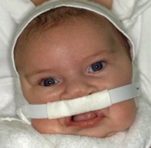
The orthodontic treatment undertaken in the first few days of life is carried out by specialist Dentists who will take a dental impression of the baby’s gum and palate. A small plate is then constructed and some strapping is applied externally to the displaced gum segments in order to realign the dental arch. The dental plate together with the strapping needs careful monitoring and adjustment to realign the displaced parts of the gum. This process is a relatively easy thing to accomplish in the first week of life when the baby’s bony structures are soft and pliable. A delay in the orthodontic treatment until 2 or 3 weeks after birth makes the job much harder if not impossible as the baby’s bones become much more rigid after the first few weeks of life. “Pre-surgical Orthopaedics”, as it is known, does not cause the baby any pain or distress. It is usual for the baby to be able to leave hospital by one week following birth. The mother will need to bring her baby back to the Dental Department at Princess Margaret Hospital for adjustments to the strapping and plate for the three month period leading up to the first operation. Mothers will be given every encouragement and assistance to manage the baby’s dental plate and strapping and most mothers become very skilful in looking after the strapping. All children managed through the PMH Cleft Lip and Palate Clinic are entitled to have their dental care undertaken at PMH. -
Surgery
The first operation is normally done at about three months of age when the cleft lip is repaired. At the same time the distortion and asymmetry of the nose is adjusted to achieve as much as possible normal nasal shape and symmetry. It is common at this first operation for some repair to be done at the front part of the roof of the mouth but not towards the back. Following the operation the baby will have stitches on the lip which remain in place for approximately five days. Once the stitches have been removed the baby is allowed to return home. It is usually not possible to continue bottle feeding for the first couple of weeks following the cleft lip repair and it is necessary therefore to feed the baby milk with a spoon at that stage. It is a good idea if the baby has been introduced to feeding with a spoon approximately one month leading up to the first operation.
The cleft in the roof of the mouth (cleft palate) is repaired at a second operation at approximately 9 months of age. On that occasion, the baby is admitted once again to hospital for approximately five days. The operation involves making an incision along the edges of the cleft and moving the tissues of the roof of the mouth allowing them to come together in the midline where they are stitched. The most important part of this repair is the soft palate which is the back of the roof of the mouth. This area is comprised of the muscles that move the soft palate to achieve normal speech. It is essential that these muscles are carefully identified, moved and stitched together in such a way that they can then function normally and move the palate properly. Without that muscle function the child will be unable to speak normally. -
Hearing
It is common for children with cleft of the palate to have some impairment of the eustachian tube function. The eustachian tube is a tunnel which extends from the back of the nose through to the middle ear and allows air to reach the middle ear. Many normal children have problems with middle ear dysfunction otherwise known at serous otitis media or glue ear. In this condition there is a build up of thick fluid in the middle ear which interferes with the function of the small bones that are critical for normal hearing.
If this build up of thick fluid in the middle ear is not corrected over a long period of time, then some permanent loss of hearing may result. Children who have serous otitis media require a small operation where tiny tubes are inserted into the eardrum allowing the fluid to drain and restoring air to the middle ear.
Children with a cleft palate are more prone to this condition and careful monitoring of hearing is required. Should the child need this operation it can usually be done as a day patient and occasionally can be combined with some other surgical procedure. In some instances, the children need several of these tubes (grommets) during the toddler years. Generally as they grow, the middle ear problems settle down and it is uncommon to require these tubes by the time the children attend school. -
Speech
The prerequisites for speech are normal hearing and normal palate function. When a child is born with a cleft palate, the muscles of the soft palate have failed to join one another in midline. During the baby’s growth in the womb the muscles are abnormally attached to adjacent bones. As a result an operation is needed, not only to close the gap in the roof of the mouth, but more importantly, to correct the misplaced soft palate muscles. Once this surgery has been completed, the muscles can then move the soft palate and direct air either through the mouth or nose to make correct speech sounds. During the toddler years the children are carefully monitored by Speech Pathologists to ensure language development and palate function are proceeding normally and if further palatal surgery is required. However, even after skilled palate repair all children require regular speech reviews at PMH at least until the age of 12 years. Depending upon their progress some children will require additional appointments and/or therapy. In some cases the PMH Speech Pathologist will liaise with the local Speech Pathology Clinic to supplement appointments.
-
Dental Care
During the time that adult and baby teeth are present together (5-12), the children are seen at regular intervals for normal maintenance of dental health. At the same time it is possible to monitor the growth of the upper jaw as this can vary considerably among individuals with a cleft of the lip and palate. An important surgical procedure carried out on children with a complete cleft of the lip and palate is an alveolar bone graft. The original surgery done of the lip and palate involved the repair of the soft tissues only. In other words, there persists a defect in the bone of the gum (alveolus) even though that bony defect is covered with gum tissues. In order to provide an optimal dental bite, an attractive cosmetic appearance and no gaps in the teeth, it is necessary to insert bone into that part of the gum where it has been missing because of the cleft. That operation is done somewhere around 10 to 12 years of age just prior to the eruption of the canine (eye tooth). Experience has shown that this is the best time to undertake a bone graft and at the operation spongy bone is harvested from the hip, either at the front or the back, and inserted into that part of the gum where the bone is missing. In the case of a child with a bilateral cleft, both sides of the cleft are bone grafted at the one operation. For this operation the young person is in hospital for approximately one week and occasionally a post operative dental may be needed. At a later date, once the canine tooth has erupted and the bone graft has stabilised, normal orthodontic treatment with braces and bands is undertaken to obtain the desired result.
Finally, in some children where the upper jaw has failed to grow adequately, an abnormal bite can result with the bottom teeth being positioned in front of the upper teeth. This can occur because of inadequate growth of the upper jaw. This may cause an incorrect bite and be unattractive. Some children can benefit substantially of both counts by jaw surgery which is best done when the children are fully grown at about 16 to 18 years of age. This small group of patients can benefit by moving the upper jaw forward surgically so that the upper teeth fit in front of the lower teeth. There are benefits both from a dental and cosmetic point of view in this small group of patients.
Cleft Palate
-
The Embryo
At about the 10th to 12th week the roof the mouth is formed in a developing foetus. The roof of the mouth forms by bone and soft tissues migrating from the sides of the mouth towards the midline where they join. That joining process commences at the front of the roof of the mouth and extends all the way back to the soft palate. Normally, the front part of the roof of the mouth is comprised of bone covered by oral lining and the back part of the roof of the mouth is soft comprising muscles covered by oral lining. It is possible, therefore, to have a failure of those tissues to join together resulting in a gap in the roof of the mouth which we call a cleft. The cleft may be confined to the soft part of the palate at the back or may extend all the way forward to the gum. Why this phenomenon occurs we do not know but there is much that can be done to correct this congenital deformity.
-
Feeding
As one might expect, where there is no intact roof of the mouth it is extremely difficult, if not impossible, for a baby to suck normally. This generally makes breastfeeding impossible but where the cleft is incomplete, or where it is not particularly wide, occasionally it may be possible for the baby to breastfeed. Where that is not possible, then feeding needs to be done with a compressible bottle, a long teat and a large hole or opening in the teat. This allows the mother and the nursing staff to squeeze the bottle and in so doing squirt milk on to the baby’s tongue, initiating the swallowing reflex and allowing the baby to feed. Usually the feeding is time consuming but with perseverance and assistance, all mothers can learn to manage this problem. Midwives who have special expertise in this area can help mothers to manage feeding. In extreme cases a baby may require a naso-gastric (stomach) tube to assist with feeding in the early stages. In some situations where the cleft is particularly wide, a dental plate may be helpful with feeding. In a wide cleft the plate may also help to prevent the tongue protruding into the cleft and this allows the gap to narrow with growth.
-
Neonatal Dental Treatment
As one might expect, where there is no intact roof of the mouth it is extremely difficult, if not impossible, for a baby to suck normally. This generally makes breastfeeding impossible but where the cleft is incomplete, or where it is not particularly wide, occasionally it may be possible for the baby to breastfeed. Where that is not possible, then feeding needs to be done with a compressible bottle, a long teat and a large hole or opening in the teat. This allows the mother and the nursing staff to squeeze the bottle and in so doing squirt milk on to the baby’s tongue, initiating the swallowing reflex and allowing the baby to feed. Usually the feeding is time consuming but with perseverance and assistance, all mothers can learn to manage this problem. Midwives who have special expertise in this area can help mothers to manage feeding. In extreme cases a baby may require a naso-gastric (stomach) tube to assist with feeding in the early stages. In some situations where the cleft is particularly wide, a dental plate may be helpful with feeding. In a wide cleft the plate may also help to prevent the tongue protruding into the cleft and this allows the gap to narrow with growth.
-
Surgery
The cleft palate is normally repaired at about 9 months of age. At this time the child needs to be in hospital for approximately five days. It is necessary in the three weeks following surgery to feed the baby with a spoon as a bottle may damage the surgical repair. It is wise for mothers to introduce spoon feeding approximately one month prior to surgery so that the baby is familiar with this method of feeding. The operation involves making incisions along the edges of the cleft and moving the tissues of the roof of the moth allowing them to come together in the midline where they are stitched. The most important part of this repair is the soft palate which is the back of the roof of the mouth. This area is comprised of the muscles that move the soft palate in normal speech. It is essential that these muscles are carefully identified, moved and stitched together in such a way that they can then function normally and move the palate properly. Without that muscle function the child will be unable to speak normally.
-
Hearing
It is common for children with cleft of the palate to have some impairment of the eustachian tube function. This eustachian tube is a tunnel which extends from the back of the nose through to the middle of the ear and allows air to reach the middle ear. Many normal children have problems with middle ear dysfunction otherwise known as serous otitis media or glue ear. In this condition there is a build up of thick fluid in the middle ear which interferes with the function of the small bones that are critical for normal hearing. Children who have serous otitis media require a small operation where tiny tubes are inserted into the eardrum allowing the fluid to drain and then the restoration of air into the middle ear.
Children with a cleft palate are more prone to this condition and careful monitoring of hearing is required. Should the child need this operation it can usually be done as a day patient and occasionally can be combined with some other surgical procedure. In some instances, the children need several of the tubes (grommets) during the toddler years. Generally as they grow, the middle ear problems settle down and it is uncommon to require such tubes by the time the children attend school. -
Speech
The prerequisites for speech are normal hearing and normal palate function. When a child is born with a cleft palate, the muscles of the soft palate have failed to join one another in midline. During the baby’s growth in the womb the muscles are abnormally attached to adjacent bones. As a result an operation is needed, not only to close the gap in the roof of the mouth, but more importantly, to correct the misplaced soft palate muscles. Once this surgery has been completed, the muscles can then move the soft palate and direct air either through the mouth or nose to make correct speech sounds. During the toddler years the children are carefully monitored by Speech Pathologists to ensure language development and palate function are proceeding normally and if further palatal surgery is required. However, even after skilled palate repair all children require regular speech reviews at PMH at least until the age of 12 years. Depending upon their progress some children will require additional appointments and/or therapy. In some cases the PMH Speech Pathologist will liaise with the local Speech Pathology Clinic to supplement appointments.